Enabling Technologies
The CaSTL Center has developed state-of-the-art instrumentation to enable the joint space-time limits of resolution relevant to chemistry. The instrumentation is available for outside researchers either as independent users or on a collaboration basis. Each instrumentation has different terms of use; please contact the faculty lead for more information.
1. Kleindiek PS8
In 2015, CaSTL and the UC Irvine user facilities jointly purchased an in situ nanomanipulation and measurement system (a Kleindiek PS8: http://www.kleindiek.com/ps8.html) for the campus scanning electron microscopes. This system enables CaSTL researchers to fabricate designer plasmonic structures that cannot be made by conventional self-assembly or lithography. The plasmonic structures are assembled by using a sharp metal tip to move nanocrystal building blocks on a substrate with ~1 nm positional control and simultaneous real-time visualization (see images and video for examples of making metamaterials such as ring-shaped oligomers and propeller-type structures incorporating particles of varying shapes, including faceted spheres, triangular prisms, and pentagonal bipyramids). We have demonstrated the ability to assemble relatively complex structures that are appropriate for single-molecule SERS and plasmon-driven chemistry. Of particular interest are structures with precisely sculpted electric and magnetic fields to enable CaSTL’s efforts in (i) time-resolved single-molecule SERS, (ii) mechanistic studies of plasmon-driven photocatalysis, (iii) chiral/magnetic plasmons, (iv) rectennas, and (v) plasmon-exciton dynamics of single nano-objects such as quantum dots. At present, the assembled structures are studied by ex situ spectroscopy. In the future, the addition of optical microscopy to the SEM will result in a true “nanoscale laboratory” that combines nanomanipulation with real-time imaging, molecular dosing, ion milling, optical spectroscopy/imaging, and electrical and other measurements inside the SEM to study the chemistry and physics of designer plasmonic structures assembled dynamically from nanoscale building blocks.
2. Femtosecond SFG microscope
Location: Ge Lab, UC Irvine
 This home-built femtosecond SFG microscope is capable of chemical imaging by time-resolving nonlinear response as well as multiplex detection in the frequency domain with submicron spatial resolution. Tunable mid-IR output from a homebuilt synchronously pumped femtosecond optical parametric oscillator (3-6 micron, 80 MHz, > 30 mW across the spectral range) is mixed with near-IR pulses from the pump oscillator (770-830 nm) onto the sample with a reflective objective inside an inverted Olympus microscope. The generated SFG signal is collected in the forward or epi direction using a photon counting photomultiplier tube. By tuning the mid-IR radiation into resonance with molecular vibrations, chemical specificity can be obtained. By controlling the time delay between the mid-IR and near-IR pulses, spatial variation of vibrational coherence time can be mapped. Alternatively, spectral resolution can be achieved by spectrally narrowing the near-IR pulses and measuring the SFG response through a spectrograph and CCD. Multimodal imaging with SHG and TPEF can also be simultaneously performed.
3. Femtosecond pump-probe scattering scanning near-field optical microscopy (s-SNOM) setup
Location: Ge Lab, UC Irvine
 In this state-of-the-art space- and time-resolved nanospectroscopy setup, a commercial platform of scattering scanning near-field microscopy (neaSNOM, neaspec GmbH) is coupled with a variety of light sources, including a He-Ne laser, a femtosecond Ti: Sapphire oscillator, and two home-built independently tunable near-IR and mid-IR optical parametric oscillators with frequency doubling options. The combination of s-SNOM technique and femtosecond laser system enables the acquisition of near-field optical images and spectra in the wavelength range from visible to mid-infrared (400 - 6000 nm) with spatial resolution of 20 nm and time resolution of 200 fs.
The setup will provide CaSTL users with unique capabilities of pseudo heterodyne near-field imaging (reflection and transmission), nano FTIR spectroscopy, and pump-probe nano-spectroscopy for understanding and exploration of nanoplasmonics and plasmon driven photocatalysis. Shown here is an example of the topography image of a Au sphere-triangle pair fabricated by nanomanipulation. The optical images taken at the 3rd harmonics of the tip tapping frequency reveal the amplitude and phase of the plasmonic near fields.
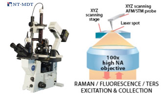
4. Integrated AFM/STM/NLO Microscope
Location: Apkarian Lab, UC Irvine
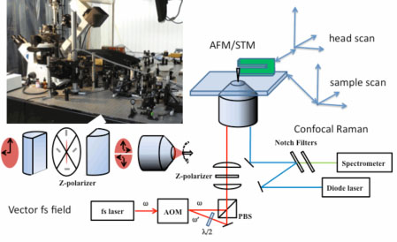 The combined AFM/STM/NLO is an inverted Olympus microscope frame integrated with a commercial AFM/STM. The system is capable to scan by sample or by head. The instrument allows for simultaneous Raman and backscatter imaging through the microscope objective, along with time-resolved measurements using vector light.
5. Ultrafast Single Molecule Imager
Location: Potma Lab, UC Irvine
 The Ultrafast Single Molecule Imager is an optical far-field microscope interfaced with an ultrafast laser light source. This system was built for the purpose to interrogate the ultrafast optical response of single molecular systems. The instrument derives its high sensitivity from several unique features. First, the laser system, a synchronously pumped femtosecond optical parametric oscillator has a very wide tunable spectral range from the visible to the mid-IR. Second, the imaging unit is equipped with three different kinds of sensitive photodectors. Besides three separate photomulitplier tubes, the microscope incorporates a low noise CCD camera and a fast photodiode for detecting modulated signals in real time. Taken together, this instrument is one of the world’s most sensitive all-optical imaging tools.
Imaging capabilities:
- Coherent anti-Stokes Raman scattering (CARS)
- Stimulated Raman scattering (SRS)
- Stimulated emission (SE)
- Second-harmonic generation (SHG) and sum frequency generation (SFG)
- Two-photon excited fluorescence (TPEF)
- Confocal reflectance
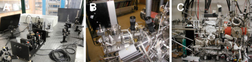
6. LT-STM with Multi-color fs Lasers
Location: Apkarian Lab, UC Irvine
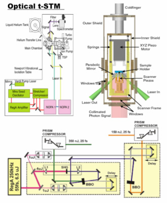 This system was designed for time-resolved STM measurements at cryogenic (5K) temperatures, and under ultrahigh vacuum (10-11 torr). A unique feature of this system is the parabolic collector (right panel), which can be aligned at cryogenic temperatures and UHV conditions to precisely match its focus with the STM tip apex. The system is coupled to a two-color fs laser, with pulse widths of 20 fs and repetition rate of 250 kHz.
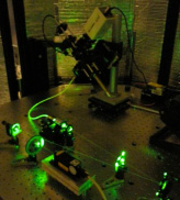
7. Sum frequency Generation Spectroscopy
Location: Ge Lab, UC Irvine
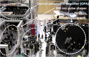 This home-built SFG spectroscopy set up is interfaced with a 90-fs, 2 kHz femtosecond laser system and consists of 1) a 100-fs OPA tunable from 3 to 10 µm; 2) a grating-slit pulse shaper for generating picosecond visible pulses; 3) a local oscillator (LO) generation unit using a LiNbO3 crystal; and 4) a focusing unit to generate SFG signals; and 5) a detection unit consisting of a spectrograph and MCT and PMT detectors. The capabilities of the set up include homodyne fs-IR/ps-vis frequency-domain experiments, heterodyne fs-IR/ps-vis frequency-domain experiments, as well as heterodyne fs-IR/fs-vis time-domain experiments.
8. fs-Å – STM
Location: Ho Lab, UC Irvine
This homebuilt system includes a femtosecond laser setup (A), a leak detector, an evaporator load lock (B), and a sample load lock (C). The laser setup has a harmonic generator (foreground in A) and a femtosecond oscillator (background in A) that provides wavelengths from 210 nm to 1040 nm, with a gap from 520 nm to 690 nm. This laser system is coupled to an adjacent low temperature STM. The evaporator load lock enables rapid turnaround of metal and molecular dosers that can be made without venting the vacuum chamber containing the STM, thus allowing rapid screening of atoms and molecules for dosing onto the sample. The sample load locks enables rapid turnaround of samples and STM tips that can be made without venting the vacuum chamber containing the STM, thus allowing rapid exchange of samples and tips.
9. TIRF/AFM Microscope
Location: Collins Lab, UC Irvine
The CaSTL TIRF-AFM system combines single molecule TIRF microscopy with a moveable, field-enhancing AFM probe in order to enable a variety of new imaging modes. On top of the light microscope, with 1.45 NA TIRF objective for single molecule fluorescence work, is mounted an AFM. It is outfitted with a high-resolution CCD camera for wide field imaging and a UV-enhanced Si avalanche photodetector for photon counting experiments.
10. Ultrafast Spontaneous and Femtosecond Stimulated Raman System
Location: Van Duyne Lab, Northwestern University
 The ultrafast system for SE-FSRS and ultrafast TERS studies consists of a high repetition-rate amplifier seeded by a broadband oscillator, which generates 8 µJ, 30 fs pulses with repetition rates from 100 to 250 kHz. Half of the amplifier output is directed to a narrow band pass filter for spontaneous and stimulated Raman pumping. Photoexcitation is performed using pulses from a visible optical parametric oscillator or fundamental doubling. A supercontinuum white light source is available for transient absorption or for stimulating femtosecond Raman spectroscopy. High-speed detection is enabled by a 1 kHz CCD (Princeton Instruments PIXIS 100F) and imaging spectrograph. This system is can perform sub-100-fs transient absorption and femtosecond stimulated Raman experiments in either a transmissive or reflective geometry.
11. Scanning Tunneling Microscope for Tip-Enhanced Raman Spectroscopy
Location: Van Duyne Lab, Northwestern University
 A home-built multi-chamber UHV system is available for atomic scale STM imaging and optical microscopy. This system prepares pristine surfaces and characterizes them at the atomic scale, with temperature control between 10 K and 400 K, with the option of performing feedback controlled nanolithography. For TERS studies, lasers at 405 nm, 532 nm, 633 nm, and 785 nm are available.
12. Ambient STM for Tip-Enhanced Raman Spectroscopy
Location: Van Duyne Lab, Northwestern University
 A custom optical microscope was fabricated for STM-TERS. Excitation at laser wavelengths given above is performed with a polarization-maintaining single-mode fiber and is focused with an achromatic lens. Inelastically scattered light from the tip-sample junction is collected through a fiber bundle and analyzed. The STM stage is controlled by the electronics system equipped with a custom approach interface. This system allows correlated TERS and STM imaging and tip-sample TERS intensity distance dependence from Au or Ag tips on any conducting substrate. Due to its ambient nature, efficient optimization of the tip preparation procedure and TERS enhancement factor can be performed on this instrument.
13. Confocal Raman Microscope
Location: Laser Spectroscopy Facility, UC Irvine
A Rennishaw InVia Cofocal Raman Microscope with excitation wavelengths 785 nm, 532 nm and 405 nm was funded by an NSF MRI-R2 Award (PI: Apkarian). The instrument is fitted with options for making a 2-dimensional Raman image of the sample, and the sample cell can be cooled to 75 K and heated to 600 ºC.
14. FEI Quanta 3D Dual-beam FIB/SEM
Location: Laboratory for Electron and X-ray Instrumentation, UC Irvine
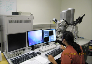 The FEI Quanta 3D FEG is a scanning electron microscope (SEM)/focused ion beam (FIB) dual-beam instrument for 2D and 3D material characterization and analysis. Various capabilities include high-resolution imaging (1.2 nm at 30 kV), low kV high contrast imaging, micromachining, Pt deposition, chemical composition analysis and mapping, crystal structure determination and orientation mapping.
15. Computation Multinode Resource
Location: The Department of Chemistry Molecular Modeling Facility, UC Irvine
The Department of Chemistry Modeling Facility is a multi-user facility that provides cutting-edge resources for performing computational simulations of chemical systems, spanning quantum-mechanical electronic structure of molecules and materials to molecular dynamics of large biomolecules and membranes. Calculations are primarily performed on a 110+ node cluster "Greenplanet", with each node having 2 quad-core 2.8Ghz Intel Xeon X5560 processors, 12-24GB 1333MHz DDR3 RAM, and 160GB-1TB local storage, and each being connected to a 40 Gigabit/s QDR Infiniband high-speed network and a 150TB disk array.
Sitemap | Privacy Policy | Term of Use
© 1996-2018 CaSTL Center or its affiliates
All Rights Reserved
Designed by WA
CaSTL Center
University of California, Irvine | 2123 Natural Sciences II | Irvine, CA 92697-2375
E-mail: CaSTL@uci.edu | Phone: (949) 824-2646 | Fax: (949) 824-8571
![]()
![]()
![]()